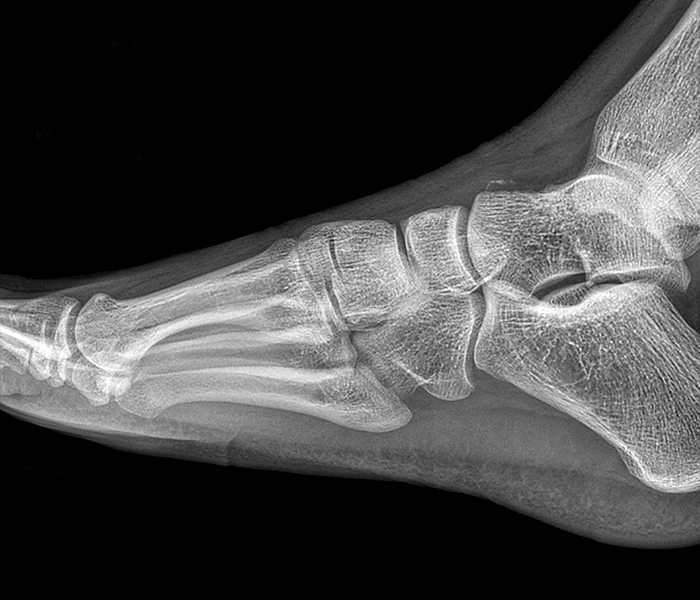
Midtarsal (Chopart) Joint Sprain

Imaging of Chopart (Midtarsal) Joint Complex: Normal Anatomy and Posttraumatic Findings
Midtarsal (Chopart) Joint Sprain
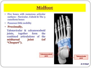
Anatomy of small joints of the foot

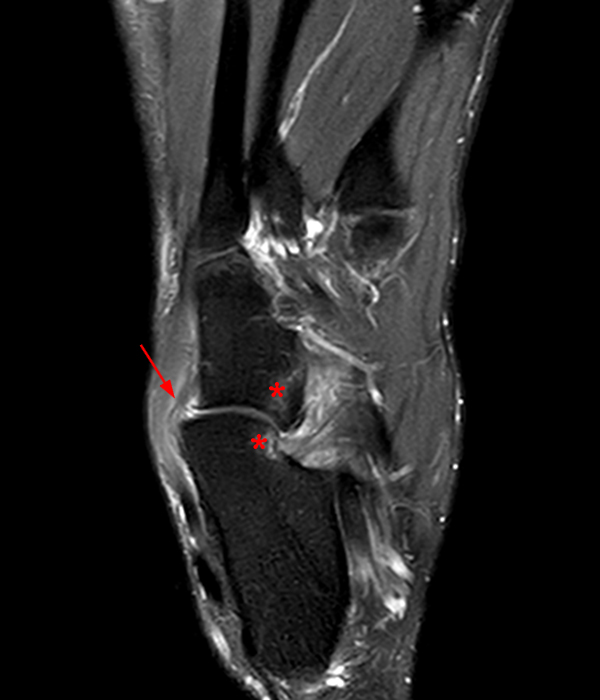
Imaging of Chopart (Midtarsal) Joint Complex: Normal Anatomy and Posttraumatic Findings

Normal Anatomy and Traumatic Injury of the Midtarsal (Chopart) Joint Complex: An Imaging Primer
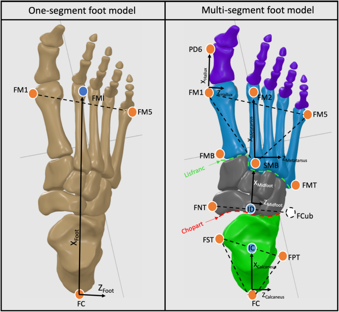
Intrinsic foot joints adapt a stabilized-resistive configuration during the stance phase, Journal of Foot and Ankle Research

Normal Anatomy and Traumatic Injury of the Midtarsal (Chopart) Joint Complex: An Imaging Primer

Imaging of Chopart (Midtarsal) Joint Complex: Normal Anatomy and Posttraumatic Findings
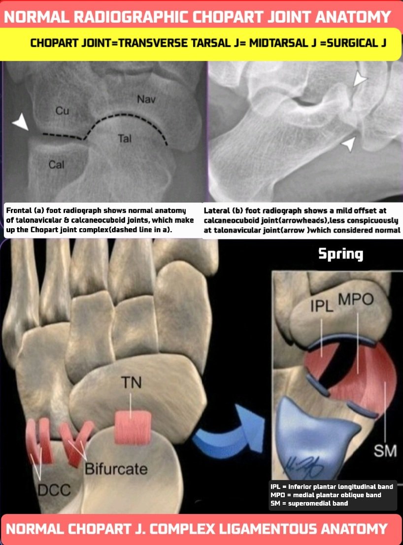
Dr.OMID BANDARCHI ,M.D. on X: 🛑CHOPART JOINT consists of the talonavicular & calcaneocuboid joints & their stabilizing lig(s): Dorsal talonavicular,Dorsal calcaneocuboid , Bifurcate,Plantar(short & long),Spring lig. A wonderful article by

Bifurcate ligament, Radiology Reference Article

Normal anatomy of the midfoot. The midfoot contains osseous