Figure 4 from The Imaging Diagnosis of Less Advanced Cases of Cardiac  Amyloidosis: The Relative Apical Sparing Pattern

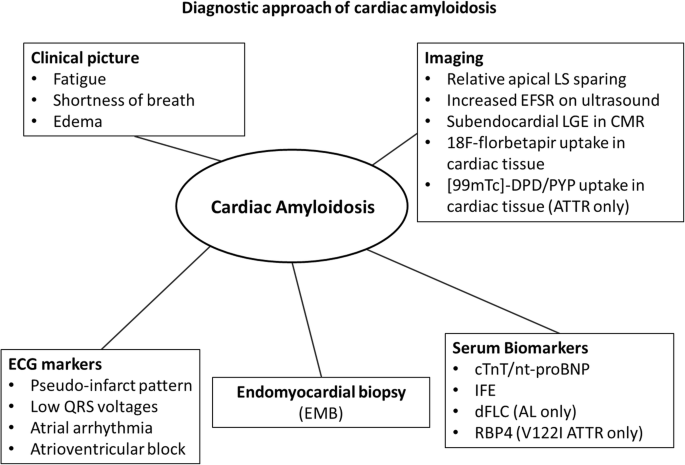
Diagnosis of cardiac amyloidosis: a systematic review on the role of imaging and biomarkers, BMC Cardiovascular Disorders

Figure 4 from The Imaging Diagnosis of Less Advanced Cases of Cardiac  Amyloidosis: The Relative Apical Sparing Pattern

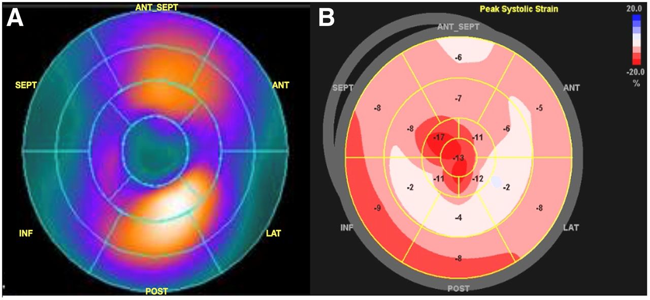
Global and Regional Variations in Transthyretin Cardiac Amyloidosis: A Comparison of Longitudinal Strain and 99mTc-Pyrophosphate Imaging

Figure 4 from The Imaging Diagnosis of Less Advanced Cases of Cardiac  Amyloidosis: The Relative Apical Sparing Pattern

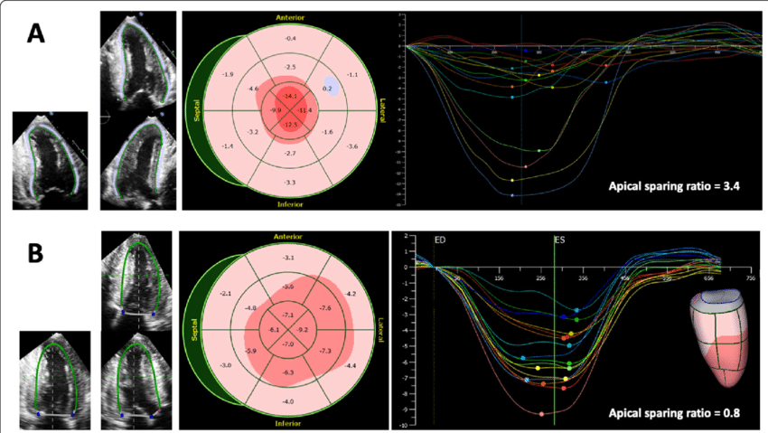
2D/3D strain analysis in a patient with cardiac amyloidosis. Upper

Figure 4 from The Imaging Diagnosis of Less Advanced Cases of Cardiac  Amyloidosis: The Relative Apical Sparing Pattern

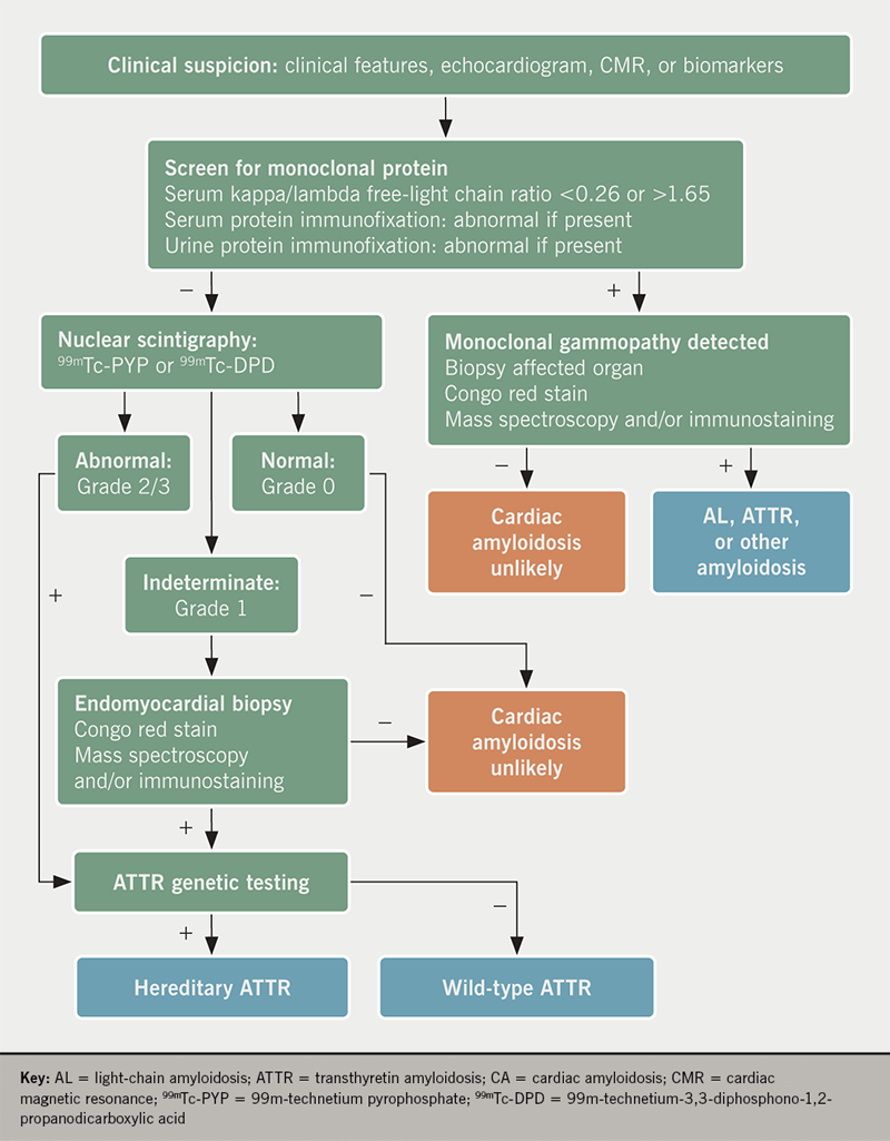
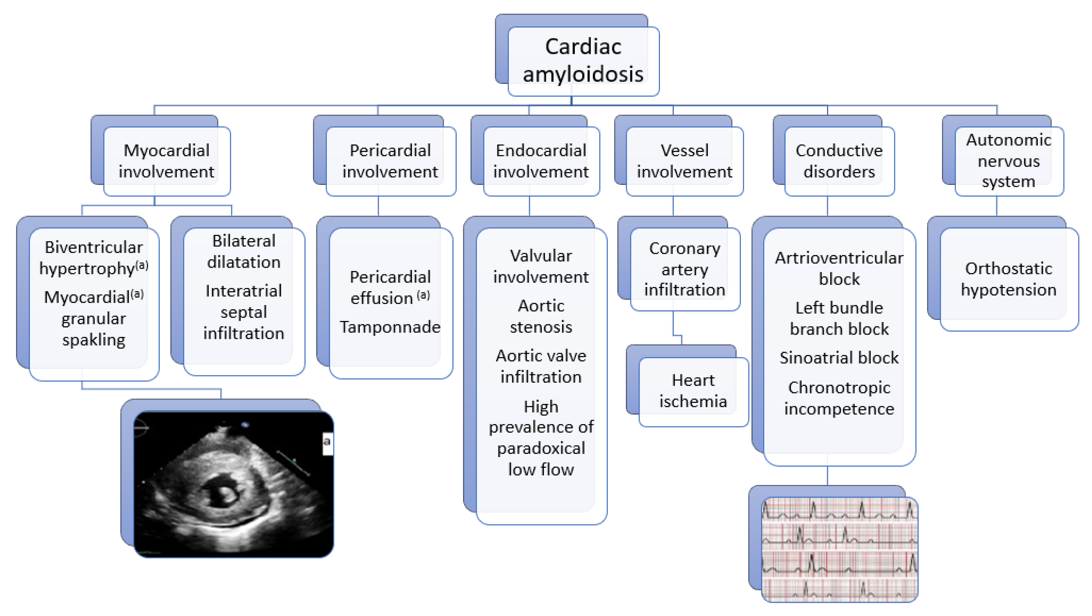
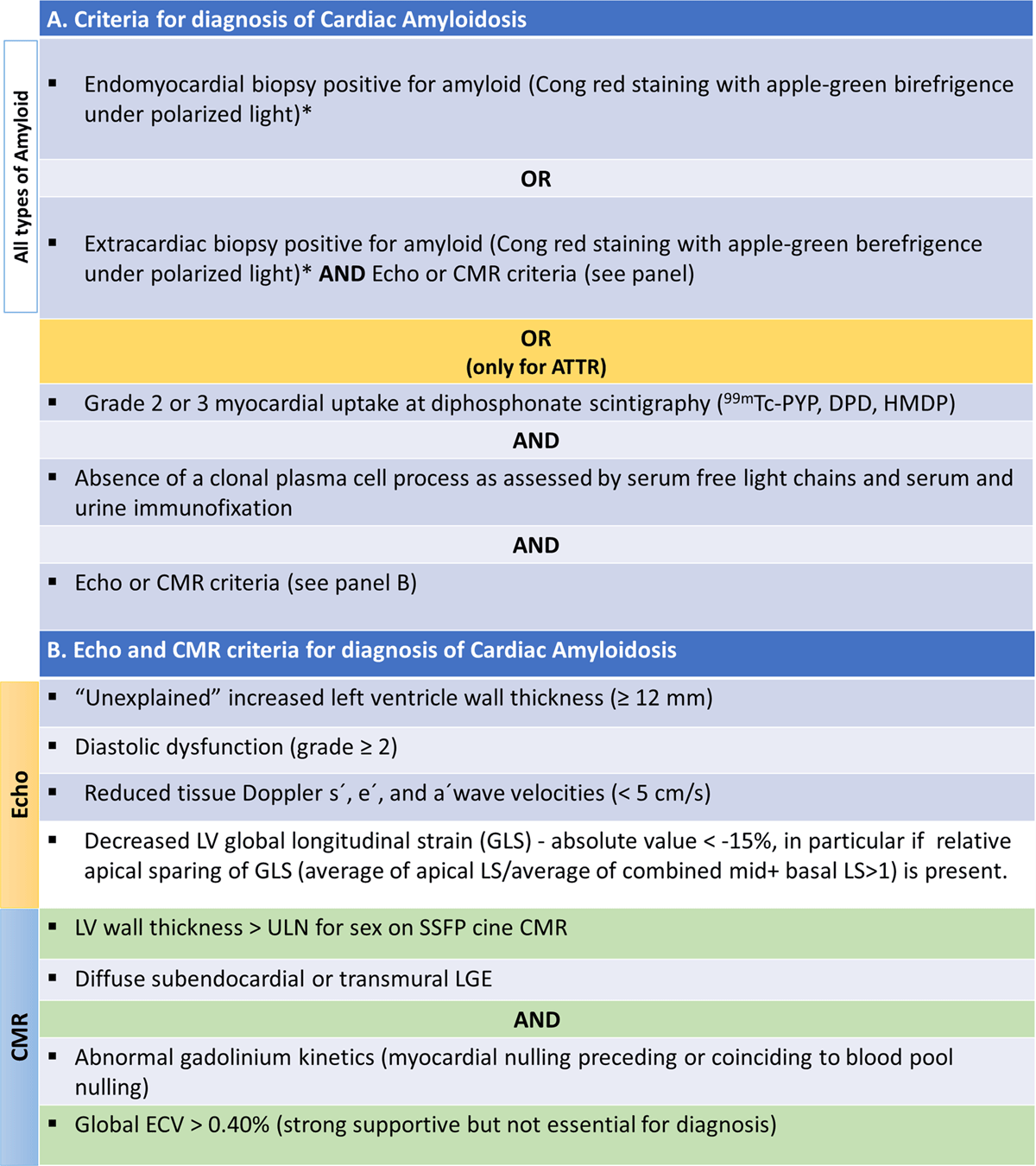
Amyloid heart disease module 1: diagnosis

Figure 4 from The Imaging Diagnosis of Less Advanced Cases of Cardiac  Amyloidosis: The Relative Apical Sparing Pattern

Biomedicines, Free Full-Text

Figure 4 from The Imaging Diagnosis of Less Advanced Cases of Cardiac  Amyloidosis: The Relative Apical Sparing Pattern

a Myocardial strain imaging by speckle tracking echocardiography of a

Figure 4 from The Imaging Diagnosis of Less Advanced Cases of Cardiac  Amyloidosis: The Relative Apical Sparing Pattern

World Heart Federation Consensus on Transthyretin Amyloidosis Cardiomyopathy (ATTR-CM) - Global Heart

Figure 4 from The Imaging Diagnosis of Less Advanced Cases of Cardiac  Amyloidosis: The Relative Apical Sparing Pattern

Relative apical sparing of longitudinal strain using two-dimensional speckle-tracking echocardiography is both sensitive and specific for the diagnosis of cardiac amyloidosis

Figure 4 from The Imaging Diagnosis of Less Advanced Cases of Cardiac  Amyloidosis: The Relative Apical Sparing Pattern

Left Ventricle Relative Apical Sparing in Cardiac Amyloidosis. - Abstract - Europe PMC